The Screening Journey: Mammograms vs. Ultrasounds
We all know regular breast cancer screenings are important, but it’s easy to become confused by the multiple tools and protocols used to detect the disease. Whether you’re new to screenings, or just need a refresher, here are some key insights to help you stay informed.
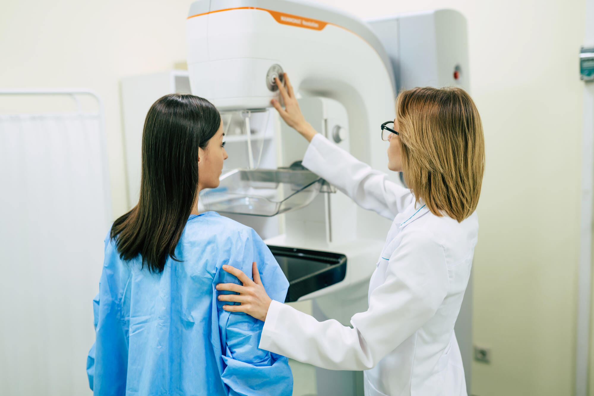
What you (still) need to know about mammograms
Mammograms continue to be the gold standard in early detection among all breast cancer screening tools. Hoag recommends that most women begin receiving annual mammograms at age 40. Decades of research have shown that they reduce mortality from breast cancer by 30-40%. Even though other tools like ultrasound and MRI have become increasingly important, the annual screening mammogram remains an essential detection tool for almost all women.
The two types of mammograms to be aware of are 2D and 3D (also known as “tomosynthesis”). While 2D was the only option prior to the FDA approval of 3D mammograms in 2011, 3D mammograms now account for 90-95% of all screening mammograms performed at Hoag. Hoag was one of the first non-academic centers in the country to perform 3D mammograms.
“You can think of a 3-D mammogram as a mini-CT scan that allows breast radiologists to view a breast in slices,” said Hoag Radiologist Nicolas von dem Bussche, MD. “Overlapping structures that impair evaluation on 2D imaging become clear on 3D. This both improves cancer detection and also decreases callbacks for benign findings.” And when it comes to which type he recommends, “it’s a no-brainer.” A 3-D mammogram improves detection for all women, but it is especially effective for woman with dense breasts.
Ultrasounds: When are they used and how are they different from mammograms?
Ultrasound uses sound waves to create images of the breast tissue. There is no radiation, and it is a useful screening and diagnostic tool that supplements mammography. For example, women who have dense tissue and/or have elevated risk (lifetime risk greater than 15%) may benefit from a screening breast ultrasound. This can either be performed at the time of the annual mammogram, or it can be performed in between annual mammograms. A screening mammogram can be performed by a technologist with a hand-held probe, but Hoag also offers automated whole breast ultrasound screening; for these exams, a series of video clips are recorded and subsequently reviewed by a breast radiologist.
“Ultrasound is also an especially useful tool to evaluate potential issues in the breast,” according to Dr. von dem Bussche. “For example, if a change is detected by the radiologist on the screening mammogram, a targeted ultrasound evaluation of the area may be recommended. Most women with a new symptom, such as a lump or pain, will also have a diagnostic mammogram performed in addition to the ultrasound, but in some situations, for example, for women under 30, or if the patient has had a recent normal mammogram, the radiologist may start with an ultrasound evaluation.”
What if a person is considered high risk?
A person’s lifetime breast cancer risk can be calculated based on a variety of factors including age, personal and family history, genetics, menstrual history, breast density, etc. A 40-year old woman with average risk will have a 12-13% lifetime chance of developing breast cancer. Women with a risk of 15-19% are considered to have intermediate risk, and a supplemental annual screening ultrasound may be recommended. Women with a lifetime risk of 20% or greater are considered high risk, and a supplemental annual screening breast MRI may be recommended.
A few terms to know:
A screening mammogram refers to a mammogram performed on a patient without symptoms or abnormalities on recent mammograms. The doctor can specify 2D or 3D, or if the patient has a preference, she can also let the technologist know prior to the exam.
A diagnostic mammogram is performed when a woman has symptoms, such as a lump, pain, or nipple discharge. This may be 2D, 3D, or a combination of both, depending on what is being evaluated. Some people with a prior low suspicion mammogram finding will continue to have diagnostic mammograms for up to 2 years. Some women with a prior history of breast cancer may also be recommended for diagnostic mammography.
A word on radiation in mammograms: When thinking about the radiation needed for an imaging exam, Dr. von dem Bussche said “different exams have completely different radiation doses and knowing the dose is all-important. It turns out that the dose for a mammogram is very low (similar to the radiation received during a transatlantic flight), and the benefits of early detection and accurate diagnosis far outweigh the risks.” Also, the difference in exposure level between 2D and 3D mammograms is negligible.
We hope this information is helpful. When it comes to navigating breast health, knowledge is critical to feeling empowered before your next appointment.